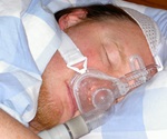
CMV Infection Linked to Increased Severity of Necrotizing Enterocolitis in Premature Babies
Researchers have discovered that infection with a common virus transmissible from mother to fetus before birth significantly exacerbates necrotizing enterocolitis (NEC), an often-fatal complication associated with premature births, in experiments conducted on mice. This study, led by investigators at Johns Hopkins Children’s Center and supported by the National Institutes of Health, advances efforts to find better treatments for NEC—a condition that is relatively rare but remains the most common emergency intestinal issue in preemies.
A report detailing this research was published on February 13th in Cellular and Molecular Gastroenterology and Hepatology. Despite its high impact, necrotizing enterocolitis is a disease that many people have never heard of. Its effect on premature infants and their families can be severe, often exacerbated by the fact that they are only introduced to this condition after their loved one has been diagnosed with it. What’s particularly surprising is our limited understanding of what actually causes NEC in the first place.
By identifying a connection between necrotizing enterocolitis and cytomegalovirus (CMV) infection, researchers have pinpointed an important trigger for this condition that could potentially save lives among premature infants who develop it. “Up to nearly 10% of premature infants develop NEC, characterized by severe inflammation in the intestinal lining which eventually leads to tissue death,” explains David Hackam, M.D., Ph.D., Garrett Family Professor of Pediatric Surgery at the Johns Hopkins University School of Medicine and surgeon-in-chief and co-director of Johns Hopkins Children’s Center. “About a third of babies with this condition ultimately die from it, and survival rates have remained unchanged over the past three decades.”
Prior animal research has suggested that NEC’s hallmark inflammation is partly due to an increased production in the immune protein called toll-like receptor 4 (TLR4), which becomes activated by specific intestinal bacteria that tend to flourish in premature infants’ digestive tracts. However, why this condition varies greatly in severity between babies and how it spreads was a mystery until now.
Considering CMV as a suspect, Hackam and his team developed a neonatal mouse model of NEC with CMV infection. They found that mice infected with CMV had significantly worse tissue damage and higher mortality rates compared to those without the virus. To uncover molecular mechanisms behind this outcome, they compared gene activity in intestines from both groups of mice.
Results indicated that CMV triggered genetic pathways promoting inflammation, disrupting metabolism, and causing more TLR4 production. Moreover, CMV damaged mitochondria—the cell’s energy factories—significantly reducing their ATP output. Additional experiments confirmed TLR4’s necessity for each effect: Mice genetically altered to produce no TLR4 in their intestines showed lower NEC severity even with CMV infection.
If future studies confirm the connection between CMV and necrotizing enterocolitis, developing treatments might include targeting this protein or administering adenosine. “When we administered adenosine to mice with both NEC and CMV, it significantly reduced the severity of the condition,” Hackam adds. The research team plans further investigation into these ideas.
Other Johns Hopkins researchers involved in this study included Chhinder P. Sodhi, Daniel Scheese, Peng Lu, Hannah Moore, Koichi Tsuboi, Cody Tragesser, Johannes Duess, Zachariah Raouf, Maame F. Sampah, Daphne Klerk, Mahmoud El Baassiri, Hee-seong Jang and Sierra Williams-McLeod.
The study was funded by grants from the National Institutes of Health (R35 GM141956 and T32 DK007713) and the Garrett Fund for the Surgical Treatment of Children at The Johns Hopkins University. Hackam, along with co-author Chhinder P. Sodhi, holds patents for developing novel agents to inhibit TLR4 for preventing or treating necrotizing enterocolitis.


